Chapter 1 : The Cells of the CNS

Brain Contents Page


Introduction to Nerve Cells and the Resting Potential

Basic Properties of Neurones

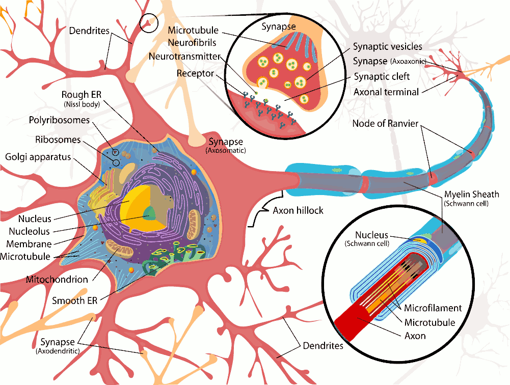
The human body is made up of trillions of cells, of which about 100 billion are nerve cells. Nerve cells or neurons, are specialized to carry electrical signals rapidly over long distances.

Neurones have specialised extensions called dendrites and axons which are involved in the collection (dendrites) and distribution (axons) of information within neural networks.

Neurones collect and integrate information in the dendrites and cell body (perikaryon) and generate action potentials at the axon hillock. The action potentials pass rapidly along the length of the axon and release neurotransmitters at the axon terminals.

Dendrites occur in large numbers, and are branches of the cell body. They receive information from other nerve cells and are not myelinated.


AXONS   Top
99

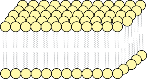
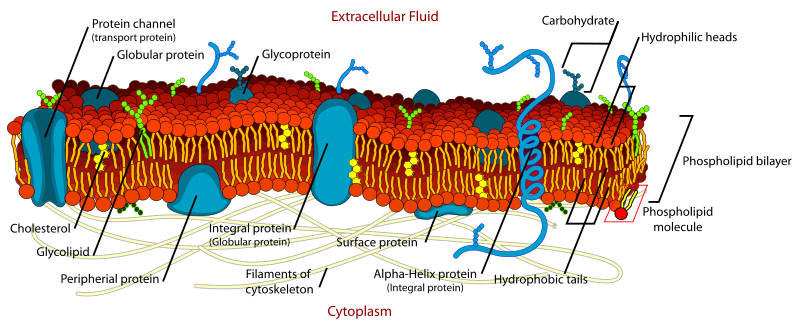
Axons, generally only one per neurone, carry information away from the cell body by means of electrical signals called action potentials that pass along the axonal membrane. This type of signalling is very fast, and is speeded up if the axon has a myelin covering. Axons usually divide into a series of branches, often at some distance from the cell body, and distribute their impulses to other cells (e.g. muscle fibres in the motor unit, or different neurones in the CNS).

Axoplasm - the cytoplasm within axons contains high [K+] and low [Na+], compared with Extracellular Fluid (ECF - high [Na+] and low [K+]). These differences in ionic concentration are crucial to one of the main functions of neurones - to conduct action potentials at high speed to the end of the axons.

Axoplasm also contains neurofilaments and neurotubules which are involved in the carriage of chemicals, vesicles and subcellular organelles to and from the nerve endings by a process called axoplasmic transport. This is a rather slow process compared with the speed of conduction of nerve impulses, and is concerned with the nutrition and connectivity of nerve endings. Substances are transported along neurotubules towards the nerve endings, and this process is called anterograde axoplasmic transport (see below)

Axons have specialized structures at their endings called synaptic boutons, which have areas of membrane specifically concerned with the release of neurotransmitters.

Neurotransmitters exert their actions on the post-synaptic cell, and can have excitatory or inhibitory effects on that cell, depending on the chemical neurotransmitter (ligand) released at the nerve ending and on the nature of the post-synaptic receptors for this ligand.

Post-synaptic cells can also communicate slowly with the pre-synaptic neurone, by releasing chemicals, such as neurotrophins, that are taken up by the nerve ending and transported slowly back to the soma, by a process is called retrograde transport.

INTRODUCTION to the ULTRASTRUCTURE of NEURONES   Top

Multipolar neurones, such as the motoneurone, can have quite complex dendritic fields, but not nearly as complex as those found in the cortex of the cerebrum and cerebellum. Others, such as the bipolar cells of the retina, are much simpler in structure.

The shape of these dendritic fields appears to be due to structural proteins in the neurofilaments that help to maintain the ultrastructure of the neurone.

The Spanish anatomist, Ramon y Cajal, uncovered some of the complexity of neurones using the Golgi staining method. The cellular theory of the brain was estabished as a result of his investigations: before his time, it was believed that the brain was simply a jelly-like organ.

Although neurones show a considerable range of structure, these variations often relate to function, and all neurones with chemical synapses function in the same basic fashion as described below.


Variety of Neuronal Structure Bipolar cells, Cerebellar Purkinje Cells, Cortical Pyramidal Cells

Top

The Neuronal Cell Membrane

The nerve cell has a body or soma, which has processes called dendrites, and an axon which is often very much longer than the diameter of the neurone. The neurones and their axons are surrounded by the nerve cell membrane, which consists of a bi-lipid layer composed of phospholipid with protein inclusions, whose functions include ion pumps and ion channels, which can transport positively or negatively charged ions across the membrane.

Lipid Bi-layer

The neuronal cell membrane is a selective semi-permeable membrane that controls the transport of materials, such as ions, into the nerve cell. It is very thin (~7 nanometres) and consists of a phospholipid bilayer which acts as an electical insulator, and appears as a double black line in electron microscopic images.

Phospholipids have water-soluble (hydrophilic) heads and fat soluble (hydrophobic) tails; these molecules are arranged in two layers with their hydrophobic tails in contact.

A consequence of this arrangement is that the water-soluble heads face the inside of the cell and the external environment of the cell. This structure is an excellent electrical insulator, and the electrical properties depend on the protein inclusions within the membrane.

Cholesterol is another lipid molecule that is an essential component of the cell membrane; it helps to provide mechanical stability and fluidity to the membrane.

Ion Channels within the Lipid Bilayer

Within the bilipid layer there are protein inclusions that have important functions that determine the electrical and other properties of the membrane. Most membrane proteins transport ions and other substances across the lipid bi-layer.

Ion movements are accomplished by the opening or closing of specific ion channels, or by active pumps that use ATP to provide energy to drive ions against their concentration gradient.

When ions move across the lipd membrane they carry charge and generate currents and transmembrane potentials. The axonal membrane is polarized at rest, and is negative inside the axon (-70 to -90 mV) because the membrane is semipermeable and potassium channels are open at rest. This potential is known as the resting membrane potential (see below).

*

Diagram showing the long lipid tails which form the hydrophobic centre of the cell membrane, and the polar ends of the hydrophilic phospholipid molecules that are in contact with the watery environment of the inside and outside of the cell.

Some protein inclusions have lipid or carbohydrate chains protruding from the membrane; these help to stabilise membrane structure, but also act as receptor molecules, e.g. for neurotransmitters. The proteins performing these functions are present in specialised structures within the cell- i.e. they are not uniformly distributed across the whole cell membrane.

Beneath the cell membrane is the cytoskeleton which gives structural support and determines the size and shape of the cell. In neurones the neurofilaments of the cytoskeleton determine the anatomy of the dendrites and the diameter of the axon.

Jerome Walker via Wiki Commons

Top

The Resting Membrane Potential

Composition of Intracellular and Extracellular Fluid

The cell membrane is the semipermeable barrier that separates the intracellular and extracellular fluids. These two compartments have every different ionic compositions.

OUTSIDE:

high [Na+] (144 mM), low [K+] (5mM) high [Cl -] (120mM)

INSIDE:

low [Na +] (15mM) and high [K+] (150mM); low [Cl-] and large [A---]

These concentration gradients are established by a combination of pumps and ion channels:

(a) the sodium-potassium pump uses energy from ATP to pump Na+ from the cell, and potassium in the opposite direction- from extracellular fluid to the interior of the cell.

(b) the cell membrane is selectively permeable to potassium ions, and potassium and diffuse freely though open channels.

Three ions of sodium move out of the cell and two potassium ions move in for every molecule of ATP consumed by the sodium-potassium pump, enduring that the internal sodium level is low, and potassium is high. Potassium ions however also redistribute themselves because of open potassium channels in the resting membrane. Whereas the membrane is rather impermeable to sodium ions.

Sodium is in high concentration in the extracellular fluid, but can't enter the cell in significant quantities even though theconcentration gradient across the membrane is high and the inside of the cell is negative, because the membrane is impermeable to sodium. Sodium channels exist in the membrane, but are closed; so no inward movement of sodium is possible in the resting neurone.

Passive movements of potassium ions

In contrast, potassium can move through open pores in the resting membrane that selectively admit potassium ions.

Potassium moves through open potassium channels, and there are two main forces that determine those movements:

(a) the concentration gradient for potassium, and

(b) the electrical gradient across the cell membrane.

The movement of an ion across the membrane depends on the concentration gradient for that ion, the electrical gradient, and the number of pores that are open to allow it to pass across the membrane.

Electrochemical Equilibrium

The concentration gradient of potassium drives it OUT of the cell; but the electrical gradient across the cell membrane causes an INward movement of potassium.

When the fluxes of potassium In and Out are equal, there is a stable equilibrium - called an Electrochemical Equilibrium; and there is a mathematical relationship between the chemical gradient and the electrical gradient in that equilibrium.

The potential at which an equilibrium is reached for any concentration gradient is called the Equlibrium Potential. The mathematical equation that describes the equilibrium potential (E) for any ion is the Nernst Equation.

E (mv)= -58 log10[ A+ inside] / [A+ outside]

Equilibrium Potentials for Na+, K+, Cl-

Ions that can diffuse freely across the membrane satisfy the Nernst Equation; these ions pass through open channels in the membrane.

Ions that do not satisfy the Nernst Equation are not freely diffusible across the membrane, either because the membrane is impermeable to that ion, or because movements of that ion are regulated by other mechanisms, such as an active pump.

For the resting membrane, the transmembrane potential in nerves is about -70mv (70mv negative inside with respect to the outside of the cell).

The Nernst Equation predicts an equilibrium potential of about -70 mv for potassium and chloride, and + 140 mv for Na+

So the resting membrane appears to be permeable to K+ and Cl- and impermeable to Na+.

The Nernst Equation predicts that an elevated external potassium concentration will affect the membrane potential. As expected raised external potassium levels cause the cell to depolarise, which is why cardiac arrhythmias are common in this condition.

Similarly, low potassium concentrations in the extracellular fluid are associated with hyperploarisation of cells, which are less excitable.

The Goldmann Equation can be used to predict the transmembrane voltage predicted by ions that are in equilibrium across the membrane (in this case, Na+, K+ and Cl-:

Intracellular Protein

Intracellular proteins are negatively charged at the normal pH of the intracellular fluid. Clearly, large molecules cannot move out of the cell, and their static charges attract positively charged ions into the cell. The charge on the intracellular proteins does not appear in the Nernst Equation, which describes the electrochemical equilibrium for potassium - this is the relationship closely resembles the electrical behaviour of the membrane.

Electrical Properties of the Cell Membrane : Resistance and Capacitance   Top

Ohm's law: describes the relationship between voltage, current and resistance: V=IR

V(millivolts)=I (milliamps) x Resistance (Ohms)

Ohms' law is a very basic equation relating a potential gradient and current flow through a Resistor. The current flow depends on the voltage gradient and the resistance (I=V/R).

For biological membranes the voltage gradient is the transmembrane potential. The resistance depends on the number if open channels and their nature; and current flow occurs when ions flow through those channels as they open or close.

Changes in membrane potential occur when ions flow through newly opened ion channels.

The next chaper deals with the Action Potential. When the neurone becomes sufficienty depolariased, a threshold is reached that activates voltage-gated sodium channels, allowing them to open. At that point some (not many!) sodium ions flow through these channels into the neurone, driven by the sodium concentration gradient and the electrical gradient. A consequence of the entry of sodium is that the interior of the neurone becomes positive with respect to the outside: this potential change is the Action Potential.

Another example is if the membrane channels for chloride suddenly open (as a result of the administration of certain pharmacological agents), negatively charged Cl- ions flow into the cell and cause the membrane potential to become more negative - i.e. the neurone is hyperpolarised.

This happens when the neurotransmitter GABA activates ligand-gated chloride GABA-A channels, allowing Cl- into the cell. (However this has nothing to do with the generation of the action potential, but illustrates another mechanism that operates in some nerve cells).

Electrical Properties of the Cell Membrane :
Capacitance

The cell membrane consists of a fatty bi-layer that causes it to have electrical capacitance. When current flows in or out of the cell, the membrane behaves like a series of capacitors (lipid bilayer) in parallel with resistors (ion channels).

This arrangement means that when current is injected (say at a single synapse) the depolarisation recorded spreads around the local membrane (according to the length constant of the membrane) and takes time to occur (depending on the time constant of the membrane).

These local electrotonic potentials are able to sum both in time and space, and characterise the local subthreshold potentials seen in neurones when current flows across the cell membrane.


Details of Membrane Properties Membrane Capacitance, Length and Time Constants, Electrotonic (local) Potentials


Chapter 1 : The Cells of the CNS

Brain Contents Page


Top

© HumanPhysiology.Academy 2014-2015